The Central Dogma
Take a paper and follow the instructions at each table.
You’re going to build your own protein and take home a glitter fortune teller!
Scroll Down for more information!
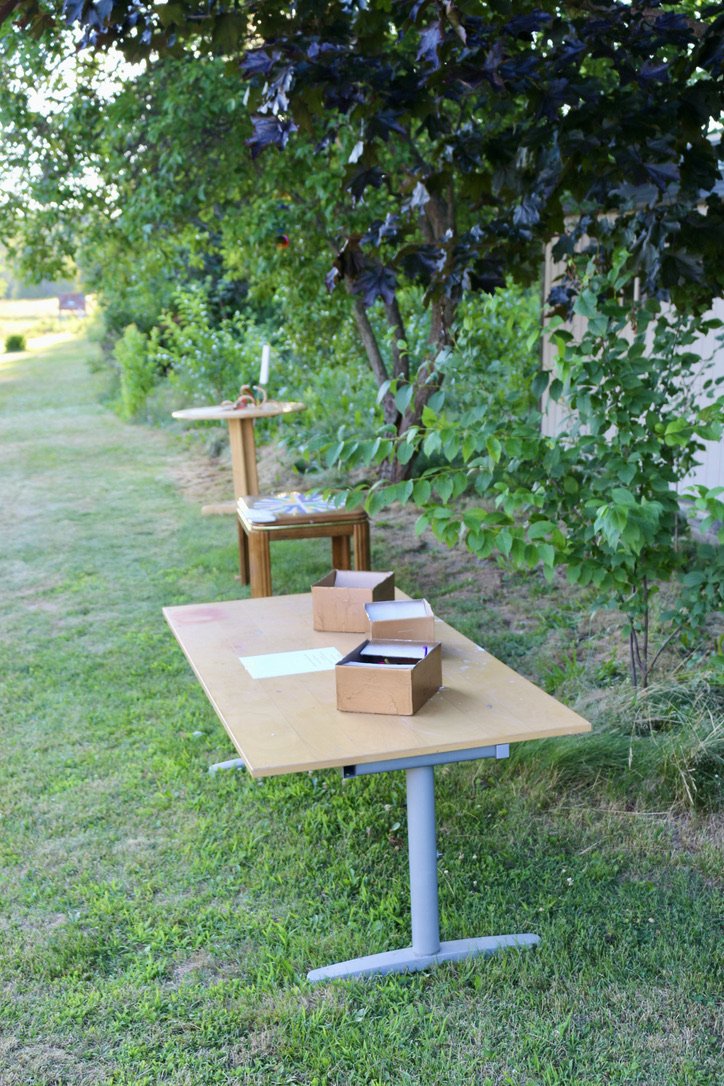
Table 1
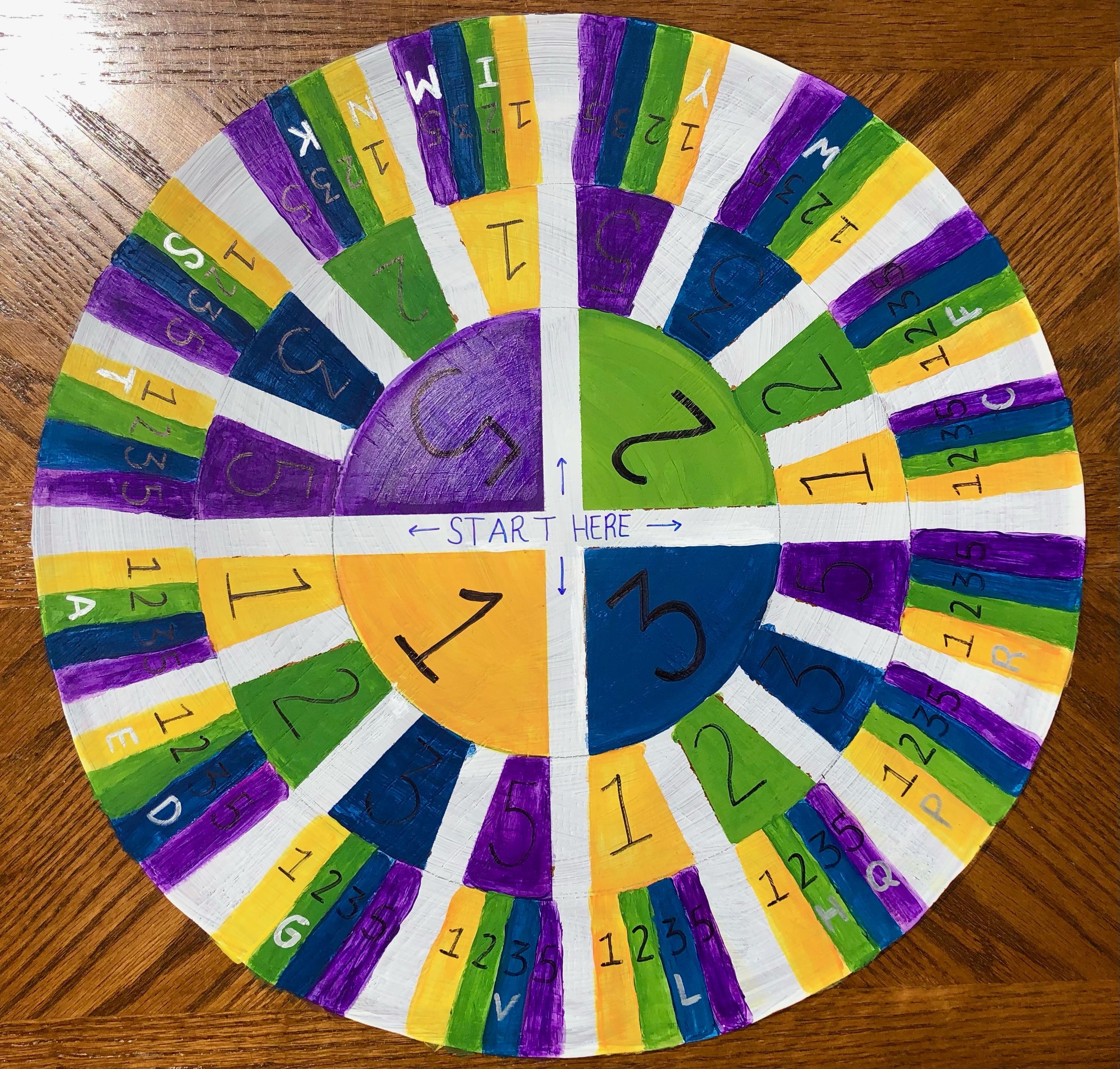
Table 2

Table 3

Fortune Teller Directions

DNA
Transcription Table
You will pick one of 3 DNA strands and record the reverse compliment on your paper
Example: DNA strand says: 1, 4, 2, 3, 1, 2, 2
You write: 3, 3, 5, 2, 3, 1, 5
(1 - 5, 2 - 3, 3 - 2, 4 -1)
Click Here for an Audio Explanation:
-
DNA, deoxyribonucleic acid, is a double stranded biological molecule, consisting of a phosphate/sugar backbone and four nitrogenous bases. These bases include: adenine, guanine, cytosine, and thymine. DNA holds all of the genetic information about all organisms, excluding the mitochondria which have their own DNA.
-
Transcription takes place in the nucleus of the cell.
-
The DNA itself, and a special protein called RNA polymerase are key. However, RNA has four bases which are matched to the DNA code, they include: adenine, guanine, cytosine, and uracil.
Are you a visual person?
Here’s a Video!

RNA
Translation Table
Using the translation table, you will turn your reverse compliment into a letter code.
You will translate every 3 numbers into one letter.
Use the translation table from the inside out.
For example: 3, 5, 1 , 1, 3, 2, 2, 2, 2
Turns into: R, G, F
-
RNA or ribonucleaic acid, is a single stranded version of DNA with its own special fourth base (That’s why there is a 5 instead of 4 as the partner!)
It holds the message that is decoded by a ribosome to make proteins.
-
Translation occurs in the ribosome.
-
RNAs and ribosomes are the key players. However, amino acids are important too. They are the letter code that equal the 3 RNA bases each.
The amino acid floats into the ribosome, where the ribosome checks if it’s the right one according to the RNA (with some error) and then clicks the amino acids together in a growing chain.
Click Here for an Audio explanation:
Are you a visual person?
Here’s a Video!

Protein
Modification Table
Looking at your letter code, you will find your modification box.
Fold your paper as instructed and then
spray with glue.
Put your fortune teller in the box, and modify away!
-
A protein is a group of compounds, strung together and folded.
They make chemical and biological changes to substrates, in order for your cell to function.
-
Protein modification occurs in thee Endoplasmic Reticulum and Golgi Apparatus. Also on thee back end of the ribosome!
-
Specialty proteins such as a hydroxlase or kinase add specific chemical groups to specific sites on the new protein.
Click Here for an Audio explanation:
Are you a visual person? Here’s a Video!
Find out about your protein!
-

Protein 1: Methyltransferase
Methyltransferases will often install a CH3 (methyl group) to a compound, DNA or other proteins.
Some methyltransferases will be modified with ubiquitination and acetylation to be more stable.
When DNA is methylated, those genes are often repressed. In other words, methylated sections of DNA are harder to transcribe.
-

Protein 2: Hydroxylase
Hydroxylases will often install an OH (alcohol group) to a compound, or other proteins.
When some hydroxylases are phosphorylated they are targeted for degradation.
Hydroxylases are responsible for modifying other proteins to make them less hydrophobic (less like fatty oils)
-

Protein 3: Fatty Acid Synthase
Fatty acid synthases are responsible for making fatty acids in the cell.
These proteins are often glycosylated.
Fatty acid synthases are mega proteins with multiple domains.
-
Impart upon the participants the Central Dogma of Biology; the pathway of a protein production follows as such: from DNA to RNA, to stitching amino acids together, to final modifications in the ER and golgi, to final product, a protein, being formed.
-
The installation will include 3 areas.
The first area will have 4 DNA models with specific sequences. The DNA sequence will be denoted with the numbers 1,2,3,4 for the four nitrogenous bases.
In this area one will match the sequence with the reverse complement (5,3,2,1) (5 will replace 4, as with thymine and uracil in DNA and RNA respectively).
Once the DNA sequence has been recorded as the reverse complement, the script will be taken to the second area.
The second area will have an RNA message decoder. As one lines up the sequence in the slots, the RNA bases (numbers) are grouped by threes.
On the table will be a chart that takes the 3 numbered sequences and turns them into letters.
Once the numbers have been turned into letters the script is taken to the third area.
The third area will have modification options such as acetylation, glycosylation, methylation, and hydroxylation. The user will match up their paper letter code with protein modification box 1,2, or 3. They will take off the layer of sticky tape, place the paper in the box and shake to modify. Each protein has a corresponding pdb file on the website to allow the user to interact with their protein.
All DNA models will be based off of real proteins.
-
http://depts.washington.edu/fhl/tidebites/Vol82/index.html
https://www.ncbi.nlm.nih.gov/Class/MLACourse/Modules/MolBioReview/central_dogma.html
https://bio.libretexts.org/Bookshelves/Introductory_and_General_Biology/Book%3A_Introductory_Biology_(CK-12)/04%3A_Molecular_Biology/4.01%3A_Central_Dogma_of_Molecular_Biology
More Advanced:
https://www.pnas.org/doi/pdf/10.1073/pnas.76.7.3164
https://pubmed.ncbi.nlm.nih.gov/26969276/
https://www.rcsb.org/structure/7S6D
https://www.ncbi.nlm.nih.gov/pmc/articles/PMC4278352/
https://pdb101.rcsb.org/motm/90
https://link.springer.com/article/10.1007/s12209-018-0151-9
https://www.ncbi.nlm.nih.gov/pmc/articles/PMC3932833/
Pdb Files for Proteins:
https://www.rcsb.org/
DNA Backbone 3D printing:
https://3dprint.nih.gov/discover/3dpx-001475
Interactive Amino Acid Codon Wheel:
https://www.sigmaaldrich.com/US/en/technical-documents/technical-article/genomics/sequencing/amino-acid-codon-wheel
-
Idea 1: To give more of an installation feel, the materials from the tables could be hung on metal structural sculptures of a nucleus, ribosome, and ER/Golgi apparatus respectively.
Idea 2: In the first area, have 3D printed models in order for the person to make an actual reverse complement of the DNA model, by snapping in the correct bases into the given 3D printed backbone.